Cardiac Image Analysis

During the past decades, Medical Imaging has played major roles in Computer Assisted Diagnosis (CAD) for cardiovascular diseases. With the recent advances in computational capability, imaging is now moving from being primarily diagnostic modality to therapeutic and interventional aid. Furthermore, with the recent developments in minimal access and Robotic Assisted Surgery (RAS), in line with the rapid emergence of biomechanic and heamodynamic modelling, the requirement for computational imaging has now reached the new height.

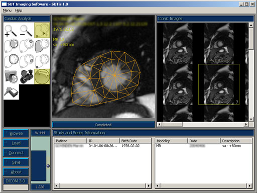
Figure 1 The SUT Imaging Suite (Freely available upon request) with a Cardiac Analysis (c) module showing an automatic ventricular segmentation procedure based on a deformable active model. The deliniated structure can then be used as a reference for many morphologic and functional assessment, e.g., cardiac output and myocardial perfusion.

 
Click [democycle.avi] for a part of segmented cardiac cycle.
Cardiac Segmentation
Contact Me
For prognosis evaluation and the assessment of the efficacy of therapeutic measures, it is neccessary to combine cardiac morphology and functions to unveil their intrinsic interactions. Conventionally, these are grossly assessed by measuring volume changes of the cardiac chambers between end diastole and systole such that global measures such as the cardiac output and ejection fraction can be calculated. To this end, one of the main objectives of the computational imaging is to present the physician with the deliniated cardiac structures, which can then be used to compute relevant indices.


With the increasing imaging capabilities in recent years, the field of cardiac segmentation and modelling is evolving rapidly. Traditional segmentation algorithm is mainly concerned with regional structure for morphological and dynamic shape analysis. Both fully automatic and manual assisted techniques have been attempted, drawing expertise primarily from Computer Vision, Digital Image Processing, and Graphics. The techniques vary from primitive region and contour based to more sophisticated model based segmentation.

Given the geometrical information of the heart, several anatomical and functional parameters may be computed. Since the reconstructed geometry contains only the description of the overall shape however the derived parameters are restricted to only that of global measures. The lack of regional correspondences has thus far limited the potential of the techniques to be successfully exploited for reliably computation of the local indices.

We have developed an approach which, on the other hand, maintain dense regional correspondence within the acquired population. As a result, the extracted geometry not only contains detailed anatomical information but also captures the intrinsic morphological variation of the shapes implied by the model. This in turn, allows phyically meaningful temporal re-sampling to assist biomecanical or heamodynamic simulation.


Journal Publications

Related References

P. Horkaew, Medical Image Computing in Cardiology, NECTEC Technical Journal, 2006 (in press).


Proceedings

P. Horkaew, Computational Diagnostic Imaging and Computer Assisted Therapeutic Intervention for Cardiovascular Diseases, in Proc. Medical Informatics, 2004, pp 175-183.